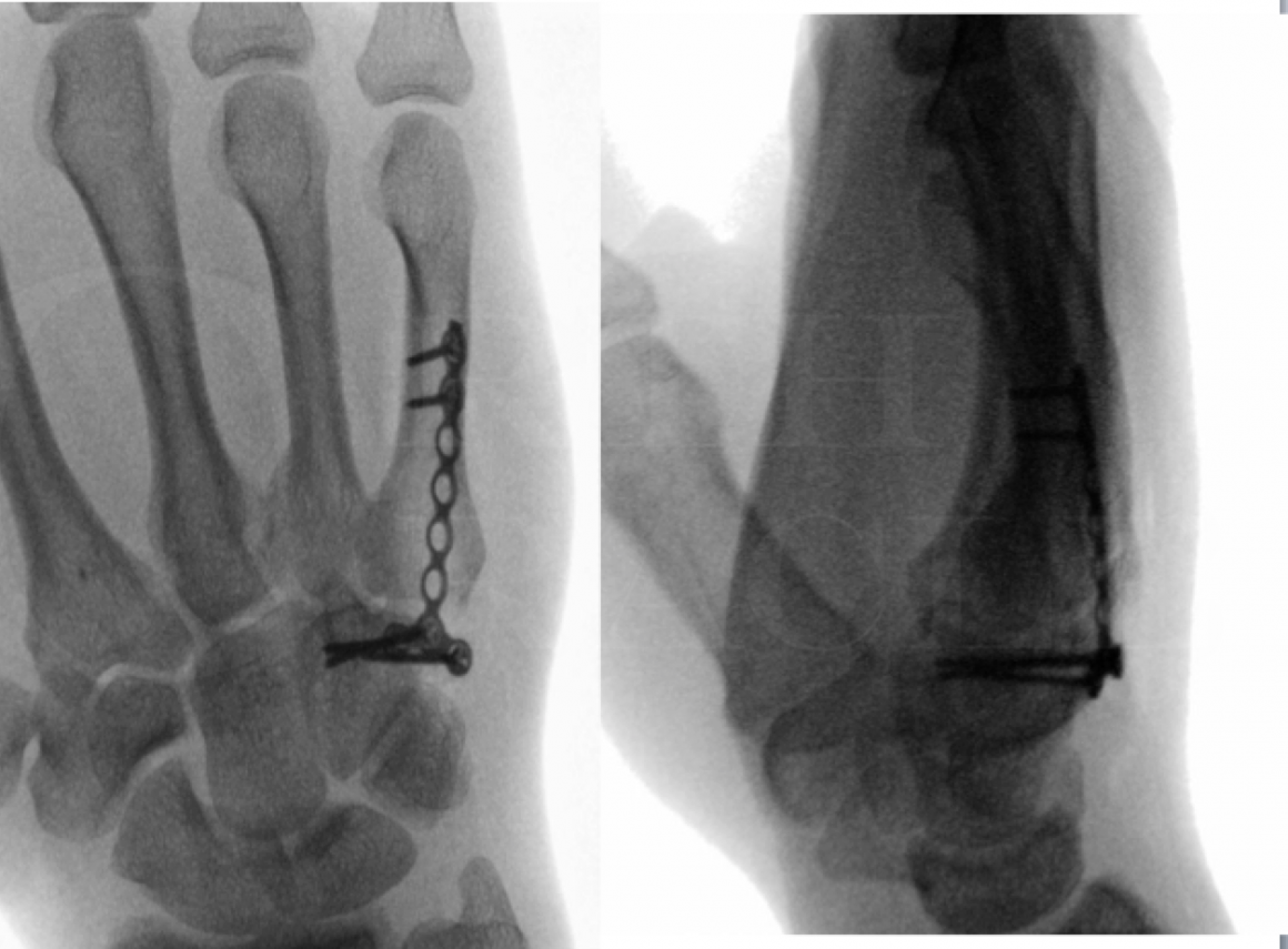
Learn the Internal fixation of 5th metacarpal neck fracture (Synthes LCP plate) surgical technique with step by step instructions on OrthOracle. Our e-learning platform contains high resolution images and a certified CME of the Internal fixation of 5th metacarpal neck fracture (Synthes LCP plate) surgical procedure.
Metacarpal neck fractures are very common in the hand. The “Boxer’s” fracture in the 5th metacarpal neck is one of the commonest injuries that present to a Hand surgeon. Most of these injuries can be treated conservatively and heal satisfactorily with no functional deficit. However, some present with rotational deformities that need correction for an optimal outcome. A handful of these also present with other concomitant injuries in the hand, which cannot undergo appropriate rehabilitation until the metacarpal neck fracture has been adequately stabilized. Many surgical techniques have been described for reduction and stabilization of this fracture with varied risks and benefits. Open fixation with plates and screws affords the most reliable reduction and a stable construct for immediate mobilization.

Indications:
Intervention with reduction and stabilization of a 5th metacarpal neck fracture is indicated in the following scenarios:
Open fractures
Fractures with rotational deformity
Comminuted and unstable fractures
Presence of associated injuries in the same hand requiring early rehabilitation
Angulation more than 70 degrees. (Although angulation at fracture site has been extensively studied, this one feature in isolation has not been found to be significant for functional outcome.)
The 2 mm locking plate (Synthes® LCP Compact Hand Set) allows for open reduction of the fracture and provides durable stability by bridging the fracture fragments. The hand can be mobilized immediately with minimal dressings.
Presentations and findings:
An axial loading force to a clenched fist causes these injuries. This produces a flexion vector on the metacarpal, which succumbs at the neck. The common mechanism is a punching injury and hence the infamous eponym of “Boxer’s fracture”. They can also coexist with high-energy injuries such as road traffic accidents – as in this case.
The patient presents with pain and swelling over the dorso-ulnar border of the hand. There is tenderness over the neck of the metacarpus and movements of the finger may be restricted with pain. A common finding is an apparent extensor lag of the small finger at the metacarpophalangeal joint. Presence of any rotational abnormality should be evaluated and documented. Ask the patient to flex all fingers together and look for any scissoring. It is important to remember that the small finger naturally curls radially and points towards the scaphoid tubercle at the wrist. Comparison can be made with the opposite uninjured hand.
Plain radiographs are essential to confirm the diagnosis and plan the management. I always request for three radiographic views of the hand– Anteroposterior, lateral and oblique. The fracture pattern and location, the displacement and the comminution are noted. Associated injuries should always be looked for and are commonly seen involving the base of the 4th and 5th metacarpals.
Alternative methods of treatment:
Conservative with closed reduction followed by plaster immobilisation – Unfortunately, this prevents early rehabilitation of the associated injuries in the hand. In addition, a plaster cast is inadequate to maintain a reduction in comminuted fractures of the neck.
Percutaneous wiring techniques– These range from longitudinal intramedullary wires, transverse intermetacarpal wires, crossed wires and Bouquet wires. Although these techniques are less invasive, they are not suitable for stabilization of comminuted fractures.
Intramedullary screw fixation – This is a relatively new technique using headless scaphoid screws to achieve stabilization and permit early rehabilitation. Comminution, however, is a contraindication for this procedure.

Informed consent is an important part of the procedure and the risks and benefits should be clearly explained to the patient. The metalwork lies in close proximity to the articular surface under the extensor tendon hood at the MCP joint. The patient should, therefore, be always counseled regarding the risk of tendon adhesions and stiffness necessitating removal of metalwork after the fracture is healed.
I prefer regional anaesthesia with axillary block for this procedure. The patient is placed supine with the limb extended on an arm table. Upper arm tourniquet is applied and inflated after exsanguination. A prescrub is performed followed by a sterile prep with Chlorhexidine. A lead hand is used to stabilize the hand. I routinely administer a single dose of antibiotics for this procedure.

The dressings are reduced in the clinic in 48-72 hours. Active mobilization exercises are commenced at this stage along with gentle passive exercises. Special emphasis is needed to mobilise the MCP joint. A splint is usually not required.
Sutures are removed in 2 weeks. Gentle routine activities of daily living can be started as soon as comfortable. Rigorous and heavy activity is avoided.
Radiographs are repeated at 6 weeks. Once the fracture healing is confirmed, aggressive passive exercises can be instituted. Activities of daily living can be increased at this stage. I advise patients against heavy activities for atleast 3 months until the fracture is consolidated.
Stiffness and tendon adhesions are a significant risk of the procedure – especially at the MCP joint. This may require a tenolysis and metalwork removal if there is no progressive improvement with therapy exercises. This secondary procedure should be delayed for atleast 3 months from the initial surgery to allow the bone to heal and consolidate.

Jahss SA: Fractures of the metacarpals: a new method of reduction and immobilization, J Bone Joint Surg Am 20:178-186, 1938. A classic original paper outlining the technique of fracture reduction that is still followed nearly 100 years later.
Wolfe, SW, Hotchkiss, RN, Pederson, WC. Metacarpal neck fracture. Green’s Oper Hand Surg. 6th edition, 2011; 241. A good overview and review of this injury along with a discussion of management including K-wire fixation. However, osteosynthesis with plates and screws is not well discussed or explained.
Poolman RW, Goslings JC, Lee J, Statius Muller M, Steller EP, Struijs PA. Conservative treatment for closed fifth (small finger) metacarpal neck fractures. The Cochrane Library. 2005 Jul 20. A Cochrane review which concluded that no single conservative method of treatment was superior to another, with all resulting in similar functional outcomes.
Padegimas EM, Warrender WJ, Jones CM, Ilyas AM. Metacarpal Neck Fractures: A Review of Surgical Indications and Techniques. Archives of trauma research. 2016 Sep;5(3). An review article that outlines the surgical indications and examines the literature on techniques of osteosynthesis. The paper concludes that internal fixation with plates and screws is the most stable biomechanics construct and is especially suitable for comminuted fractures although carrying higher risks of complications.


