Learn the Comminuted clavicle fracture – internal fixation with Stryker Variax 2 locking plate surgical technique with step by step instructions on OrthOracle. Our e-learning platform contains high resolution images and a certified CME of the Comminuted clavicle fracture – internal fixation with Stryker Variax 2 locking plate surgical procedure.
Clavicle fractures are common accounting for around 4% of all fractures and up to 44% of fractures of the shoulder girdle of which the middle third is by far the most common site. The management of such injuries can be difficult and outcome can be unsatisfactory. There has been reported non-union rate of displaced mid-shaft clavicle fractures of between 15 and 20%. This can be reduced significantly with surgical intervention. Whilst there is risk and some potential complications with any surgical intervention the published results demonstrate that plate and screw fixation can be performed safely to give a good outcome with improved patient satisfaction and a reduction in the non-union rate compared to conservatively managed fractures.
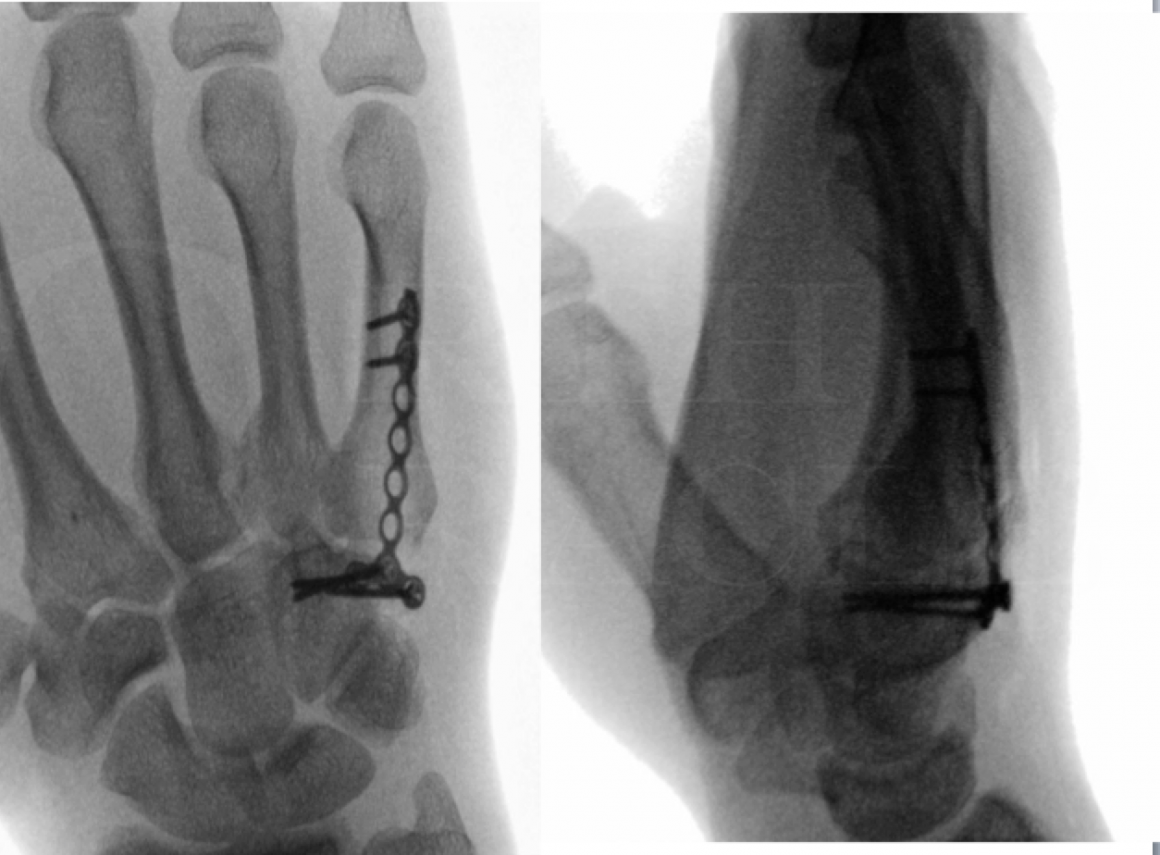
There are many implant companies with clavicle specific plates. Described here is the use of the Stryker VariAx 2 clavicle locking plate system. These contoured and sided plates are available for lateral injuries, superior shaft fractures and for use along the anterior face of the clavicle. The plates and screws are made of different grades of titanium allowing the harder locking screws to cut their own thread within the softer material of the plate and their position can be adjusted up to three times. Screws are available in sizes 3.5 and 2.7 in both non-locking and locked varieties. The clavicle set also provides useful instruments to facilitate exposure with periosteal elevation, fracture reduction in terms of clamps and wires as well as plate holding clamps.

INDICATIONS
Surgical fixation of clavicle fractures is a source of recent significant debate, research and ongoing discussion within the shoulder & trauma surgery communities.
The indications for internal fixation of a clavicle fracture are relative. The vast majority of such fractures will unite with conservative measures. The advent of contoured fracture specific locking plates have been useful in the management of such injuries. Several recent high profile publications have looked at the outcomes from randomised trials of fixation of these. I believe that patients should be considered for surgery in the presence of a comminuted , displaced and mid-shaft fracture such as is presented here. One absolute indication though for surgery would be an open injury with penetration of the skin. Tenting or threat to the skin is a relative indication as is significant shortening of the shoulder girdle due to overlap of the fracture fragments.
In my unit we adopt a policy of initial conservative management until the acute injury has settled and then a fracture clinic review by senior trauma or shoulder surgeon to assess progress within the first two weeks. In some patients the initial pain has settled significantly such that they are quite happy to pursue a non-operative course. A second group of patients will be struggling with pain and deformity in which case we offer surgical intervention.
SYMPTOMS & EXAMINATION
It is important to ascertain that this was a normal shoulder prior to the injury . The fracture usually results from a fall onto or direct blow to the shoulder girdle. This often happens as a result of sport or leisure activities particularly mountain bikes or contact collision team ball sport such as rugby. Patients present with pain and associated swelling around the region of the clavicle. Initially they will be reluctant to move the shoulder however it is important to ascertain whether there is still glenohumeral movement. Care must be taken to avoid assuming that the obvious fracture in the clavicle is the only injury in the shoulder. Previous shoulder trauma or indeed intervention should be ascertained. At the initial presentation clinical assessment of the rest of the shoulder girdle can be very difficult due to the acute pain. Important features to assess and document are the neurovascular status of the upper limb.
IMAGING
2 view plain radiograph X-rays are mandatory. Cross sectional imaging may be indicated if there is concern about a fracture towards the medial end and the sternoclavicular joint as this is notoriously difficult to image with plain X-rays. CT scans or indeed MRI scans can be useful looking for occult injuries around the shoulder and should be assessed on a case-by-case basis. Should there be any concerns as to the vascular status of the upper limb then close liaison with local vascular surgery colleagues is important and consideration should be given to angiography or contrast imaging.
ALTERNATIVE OPERATIVE TREATMENT
The technique described here is a superior contoured plate but alternatives would be anterior plating or an intramedullary device.
NON-OPERATIVE MANAGEMENT
It would be quite acceptable to propose non-operative treatment in such a patient with immobilisation in a sling or figure of 8 bandage or brace allowing underarm hygiene and encouraging elbow wrist and hand movements. Pendular shoulder exercises should be started early and then as pain settles active assisted shoulder movements can be commence. Most patient with such an injury will require sling immobilisation for the best part of the first four weeks and may struggle to be free from the sling until six weeks. Continued conservative management with graduated physiotherapy rehabilitation to concentrate on regaining range of shoulder motion prior to strengthening is well established.
CONTRAINDICATIONS
Patients’ co-morbidities and medical state should be assessed as to whether they are fit enough for surgery under general anaesthetic. Patients should be compliant with a post-operative regime as described. The state of the skin should be carefully assessed in the initial period to ensure that there is no soft tissue and skin abrasions over the site of surgical incision as this would be a relative contraindication to immediate surgery. Often surgery will be postponed or delayed due to the presence of fresh skin contusions or abrasions.

In the semi-sitting or beach chair position the patient is secured with their head on the head ring of a shoulder specific table attachment. Intravenous antibiotics are administered by the anaesthetist. Intermittent calf compression is used for thromboembolic prophylaxis during the procedure. A narrow moveable arm table is used on the operated side to rest the arm in a comfortable position. The shoulder should be placed on to a radiolucent part of the operating table to allow easy access for image intensifier and images during surgery or for checking the final position of fracture reduction and metalwork.


Prior to discharge from hospital the patient is instructed in underarm hygiene and mobilisation of elbow, wrist and hand. Active shoulder mobilisation is permitted avoiding elevation of arm above shoulder height for first three weeks. Review in fracture clinic at two weeks allows inspection of the wound following removal of Steristrips. The free ends of the absorbable Monocryl suture are then trimmed at skin level. Check X-rays are taken at this stage. The patient is allowed to wean from the sling as comfort allows over the next two weeks, increasing active shoulder mobilisation but avoiding lifting and resistance until comfort at four weeks. Discard sling at four weeks. Continue with graduated active shoulder mobilisation with physiotherapy instruction until eight weeks. If X-rays are satisfactory at eight weeks can return to full activities as comfort allows.

Nonoperative treatment compared with plate fixation of displaced midshaft clavicular fractures. A multicenter, randomized clinical trial. Canadian Orthopaedic Trauma Society. J Bone Joint Surg [Am] 2007;89-A:1-10
Locked intramedullary fixation versus plating for displaced and shortened mid-shaft clavicle fractures: a randomized clinical trial. Ferran NA, Hodgson P, Vannet N, Williams R, Evans RO. J Shoulder Elbow Surg 2010; 19:783-789
Open reduction and plate fixation versus nonoperative treatment for displaced mid-shaft clavicular fractures: a multicenter, randomized, controlled trial. Robinson CM, Goudie EB, Murray IR et al. J Bone Joint Surg [Am] 2013;95-A:1576-1584
Early versus delayed operative intervention in displaced clavicle fractures. Das A, Rollins KE, Elliott K, Johnston P, van Rensburg L, Tytherleigh-Strong GM, Ollivere BJ. J Orthop Trauma 2014;28:119-123
The Clavicle Trial:a multicenter randomized controlled trial comparing operative with nonoperative treatment of displaced midshaft clavicle fractures. Ahrens PM, Garlick NI, Barber J, Tims EM, Clavicle Trial Collaborative Group. J Bone Joint Surg [Am] 2017; 99-A:1345-1354