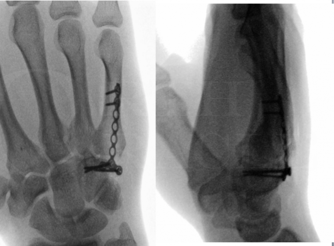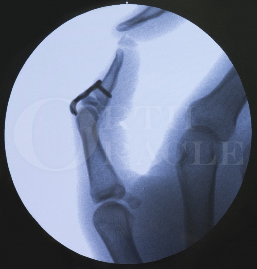
Mallet injuries are amongst the most common injuries involving the fingers of the hand. These are avulsions of the extensor tendon from their attachment at the dorsal base of the terminal phalanx. Some of these avulsion injuries may have a bony component of variable size. These seemingly simple injuries require a long and tedious management with less than satisfactory outcomes. A majority of them can be treated conservatively in a mallet splint. However, some of the displaced bony injuries require a reduction of the Distal Interphalangeal Joint and a stabilization of the fractured fragment. A hook plate is an elegant technique to facilitate this. It protects the avulsed bony piece from fragmentation with metalwork insertion, and provides a stable construct for reduction and fixation.

Doyle had classified mallet finger injuries into 4 types:
Closed injury with/without small avulsion fracture
Open injury with tendon laceration
Open injury with tissue loss
Mallet fracture
Wehbe & Schneider have further detailed the classification of bony mallet fractures based on the size of the fragment and any subluxation of the DIP joint. I consider ORIF with a hook plate when there is a displaced fracture involving >40% joint surface. I believe that the remaining articular surface is otherwise insufficient to provide a stable reduction of the DIPJ, and is therefore at a higher risk of displacement and subluxation.
Contraindications:
Injury more than 4-5 weeks is classed as a chronic mallet deformity and is not amenable to surgical fixation. A trial of splint can be tried, but these patients may require future tendon reconstruction or DIPJ fusion depending on their long-term symptoms.
Presentation and findings:
Mallet injuries result when there is an external forced hyperflexion or axial loading to an actively extended fingertip. Common mechanisms include snagging the extended finger in a bed sheet or a blunt impact to the fingertip while trying to catch a ball. This particular patient was involved in a road accident and the bony mallet injury was one of a spectrum of injuries he sustained.
In a closed injury, patients present with a typical deformity with a drooping tip of the finger at the DIPJ– the so called “mallet deformity”. They lack active extension at this joint (extensor lag). Active flexion, however, remains well maintained. There is usually swelling and some associated pain on the dorsum of the DIPJ. Nail complex is uninvolved in an isolated injury.
Investigations with plain radiographs are essential to confirm any bony fragment and its displacement. The lateral view provides the most useful information. The X-rays will also help identify any subluxation of the joint. Further imaging is usually not required to plan the management.
Alternative methods:
Conservative with splinting – Numerous authors have shown that most such injuries can be managed reliably with splintage. Even the specific type of splint has no bearing on the final outcome. However, there are no meaningful trials comparing surgery with splint in displaced bony fragments that involve a significant proportion of the articular surface – as shown in this particular case. I have already explained my reasoning for surgical intervention in such scenarios.
Axial wiring – Although this is a technically simpler procedure to perform, it does not address the displaced bony fragment. In addition, it necessitates immobilization of the DIP joint until the wire is removed in 6-8 weeks.
Ishiguro wiring – This technique stabilizes the bony fragment with an additional dorsal blocking wire. However, reduction of the fragment may be less than optimal. This procedure also requires immobilization of the joint until removal of the K wires.
Interfragmentary screw fixation – The bony fragment in mallet injuries is usually very small and attempts at insertion of lag screws carries significant risk of comminution of the fragment. The hook plate avoids this by buttressing the fragment without inserting any metalwork through it.

Informed consent is an important part of the procedure and the risks and benefits should be clearly explained to the patient. The metalwork lies in the subcutaneous tissue and often requires removal at a later stage. The distal edge of the plate lies close to or under the germinal matrix of the nailbed and may result in some nail growth abnormalities and deformities. Some persistent stiffness in flexion and extensor lag at the DIP joint, are unfortunately related to the injury and occur with all available options of management. Alternative methods should always be deliberated and an opportunity given to the patient to choose a nonsurgical option.
The procedure can be performed under local ring block anaesthesia – especially in the long fingers of the hand. I prefer a regional axillary block for the thumb. A digital tourniquet allows for a bloodless field and adequate visualization. Intra-op imaging should be planned and arranged. The patient is placed supine with the arm outstretched on a hand table. A “lead hand” may be used to stabilize the hand. I routinely administer a single dose of antibiotics for this procedure because it involves implantation of metalwork.


Dressings are reduced in 2-4 days. A light dressing allows commencement of early therapy. One of the advantages of this procedure is that the patient can start mobilization exercises immediately. Active flexion at the DIP joint produces a tension band effect at the fracture site – allowing compression and promoting healing. An intermittent mallet splint is provided to the patient for support during this period.
Sutures are removed at 2 weeks, at which point the wound is left open and scar massage started.
Mallet splint is weaned off in 6-8 weeks. Check X-rays are done at this stage. Healing of the fracture is confirmed before commencing unprotected mobilisation.
As the metalwork is subcutaneous, I find that it requires removal in most cases. However, I prefer to withhold the removal of metalwork for atleast 3 months.

Botero SS, Diaz JJH, Benaida A, Collon S, Facca S and Liverneaux PA. Review of acute traumatic closed mallet finger injuries in adults. Arch Plast Surg. 2016, 43: 134-44. This is an excellent review article detailing the current concepts of pathogenesis, classification and management principles of a mallet injury. They added their own modification to the classification system.
Doyle JR: Extensor tendons—acute injuries. In Green D (ed): Operative Hand Surgery, 4th ed, New York, Churchill Livingstone, 1999: 195-198. Classification of mallet injuries, which has then been reproduced in later editions as well.
Handoll HH, Vaghela MV: Interventions for treating mallet finger injuries, Cochrane Database Syst Rev (3):CD004574, 2004. The review concluded that there is insufficient evidence to support operative over non-operative treatment modalities. However, it noted that there weren’t sufficient appropriate trials to make any robust recommendations.
Teoh LC, Lee JY. Mallet fractures: a novel approach to internal fixation using a hook plate. The Journal of Hand Surgery: British & European Volume. 2007 Feb 28;32(1):24-30. This was the first description of the hook plate in a consecutive series of 9 patients. The authors detailed the technique and reported good outcomes.
Acar MA, Güzel Y, Güleç A, Üzer G, Elmadağ M. Clinical comparison of hook plate fixation versus extension block pinning for bony mallet finger: A retrospective comparison study. J Hand Surg Eur. 2015, 40: 832-9. The authors concluded that open surgery with hook plate resulted in better fracture reduction and an earlier return to function. The final extensor lag and stiffness were not significantly different one either group.